: +91(0)431-2760433

: info@krs-ipl.com

Portal Dosimetry System
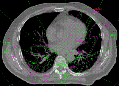
Portal Dosimetry System for true 3D IMRT and IMAT dose verification and dose reconstruction with absolute dose using available EPID, ion chamber array, or diode array for conventional linacs, and the TomoTherapy detector array for TomoTherapy:

  • Measure the radiation fields pre-treatment.
  • OR measure the fields during treatment for exit (transit) dose reconstruction.
  • Using measurement is working backwards to compute the dose to the patient.
  • Compare to the plan dose.

  • The program uses a pencil beam algorithm. The pencil beam is developed from measured beam data and Monte Carlo calculated kernels. A single poly-energetic kernel is used to represent the pencil. There were two considerations for making this choice. First we would like the algorithm to be as fast as possible. Calculating each energy separately with a spectrum and mono-energetic kernels would reduce the speed significantly. It is our philosophy that if greater accuracy is desired, one may as well use a Monte Carlo algorithm, so that there is a fast algorithm for quick results and a slow one for greater accuracy. Second, for the Dosimetry Check application, we have to consider the input information.

    Technical Specs:

  • 3D In-Vivo Patient Exit Dosimetry
  • Transit Dose Export to any 3rd Party Software.
  • Uses an independent 3D dose calculation algorithm to calculate dose
  • Can perform secondary check like any other Mu Check software.
  • Directly demonstrates the dose to the patient using the Volumetric CT Data Set
  • Can use customers measured beam data to match TPS output
  • User defined dose matrix
  • Calculates dose using the entire CT Data set
  • Uses EPID as the measurement device for both absolute calibration and intensity maps
  • No additional hardware requirements
  • Provides 0.25mm sample resolution
  • Displays composite dose for all beams
  • Displays dose of each single beam
  • Displays dose in a 3D Dose cloud
  • Displays dose in the Coronal, Sagital or Axial planes
  • Can reformat CT Image in any plane and display dose
  • Calculate and display 1D profiles in any plane through the CT data set
  • Displays reconstructed calculated dose and planning system dose in any generated CT image
  • Displays 3D volumetric comparative Dose Volume Histograms
  • Displays 3D Gamma Volume Histograms Structure by Structure
  • Imports TPS Calculation Points (Dicom RT)
  • User can add additional 3D points in DC
  • Calculated points anywhere in the CT data set and displays calculated vs. measured
  • User defined inhomogeneous correction
  • Provides MLC QA tools using the EPID: distance function and profile function on EPID image.
  • Measure distance function to determine leaf position
  • Deconvolution Algorithm to remove scatter from the EPID
  • Contouring tools